

A single misinterpretation of a monoisotopic mass peak can invalidate months of rigorous laboratory research. In the precise field of biotechnology, the inherent complexity of interpreting mass spectrometry data for peptides often creates an analytical bottleneck that threatens the integrity of your experimental data. You likely recognise that distinguishing between a genuine peptide signal and a spectral artifact is not merely a technical step; it's a critical safeguard for your project's validity. Confusion between monoisotopic and average mass, or the presence of unexpected sodium and potassium adducts, can easily obscure the truth of your results.
This protocol provides a definitive framework to master systematic deconvolution and ensure absolute sequence verification in your research. By refining your approach to spectral analysis, you'll gain the clarity needed to identify subtle impurities and maintain the highest standards of quality assurance. We will examine the nuances of isotope distribution, the identification of common spectral contaminants, and the implementation of standardised documentation for professional laboratory records. This structured methodology ensures that your findings are supported by the quiet confidence of technical precision and scientific rigor.
Key Takeaways
- Learn a systematic protocol for interpreting mass spectrometry data for peptides to ensure every research sequence is verified with absolute precision and technical rigour.
- Master the deconvolution of complex m/z spectra by accurately identifying multiple charge states and distinguishing between monoisotopic and average mass peaks.
- Utilise the Roepstorff-Fohlman nomenclature to map fragmentation ions, allowing for the definitive reconstruction of peptide sequences through tandem mass spectrometry.
- Minimise spectral noise by isolating common cationic adducts such as sodium and potassium, ensuring that potential impurities are correctly identified during quality control.
- Standardise laboratory documentation by integrating raw spectral data with Certificates of Analysis to maintain a meticulous audit trail for high-purity research compounds.
Establishing Scientific Rigour in Peptide Analysis
Mass spectrometry serves as the definitive gatekeeper for laboratory integrity. In modern research, the process of interpreting mass spectrometry data for peptides ensures that a sequence is not merely present, but is structurally correct and free from synthesis by-products. This analytical rigour is particularly vital for high-potency compounds such as retatrutide, where even minor sequence errors or truncated impurities can drastically alter experimental outcomes. Without precise verification, the validity of a study's entire dataset remains in question.
While simple identification confirms the molecular weight, quantitative purity analysis requires a deeper investigation of the spectral baseline. The clinical workflow follows a structured sequence:
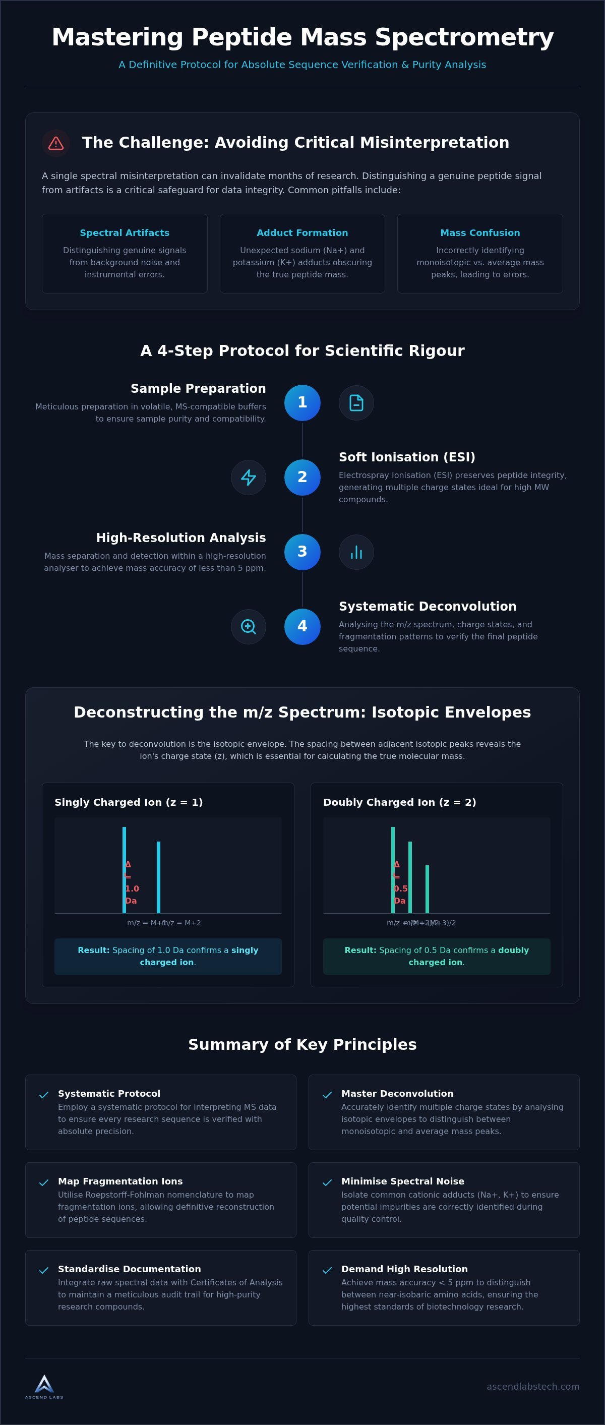
- Meticulous sample preparation in volatile, MS-compatible buffers.
- Soft ionisation to preserve the structural integrity of the peptide.
- Mass separation and detection within a high-resolution analyser.
- Systematic deconvolution of raw data for sequence verification.
This methodical approach allows researchers to validate the Ascend Labs peptide catalogue with the high degree of transparency that advanced practitioners demand. It's the difference between assuming quality and proving it through empirical data.
The Necessity of High-Resolution Spectrometry
Distinguishing between nominal mass and accurate mass is the first step in establishing scientific truth. Nominal mass provides a rounded integer, while accurate mass provides the decimal precision necessary to calculate a compound's elemental composition. In a laboratory setting, mass accuracy is measured in parts per million (ppm). Achieving a mass accuracy of less than 5 ppm is essential when distinguishing between near-isobaric amino acids, such as glutamine and lysine, which differ by only 0.036 Da. Rigorous spectral validation is the only way to ensure that research-grade peptides meet the exacting standards of contemporary biotechnology.
Ionisation Techniques for Peptide Research
The choice of ionisation dictates the complexity of the final spectrum. Electrospray Ionisation (ESI) is generally preferred for fragile metabolic research compounds because it's a "soft" technique that minimises fragmentation during the initial phase. Unlike Matrix-Assisted Laser Desorption/Ionisation (MALDI), which often produces singly charged ions, ESI frequently generates multiple charge states. This characteristic is advantageous for interpreting mass spectrometry data for peptides of higher molecular weights. Understanding protein mass spectrometry techniques is fundamental to selecting the right ionisation mode, as the resulting ion distribution directly impacts the ease of deconvolution and sequence verification.
Deconstructing the m/z Spectrum and Charge States
The m/z ratio is the fundamental coordinate of every mass spectrum. It represents the mass of a peptide divided by its net charge. While the concept is straightforward, the complexity of interpreting mass spectrometry data for peptides arises from the fact that most compounds don't exist as simple, singly charged ions. In electrospray ionisation, peptides frequently acquire multiple protons, resulting in a series of peaks that represent different charge states ($z = 1, 2, 3+$) of the same molecule. Standardised Peptide Mass Spectral Libraries provide the benchmarks required to verify these patterns against known sequences.
Accurate deconvolution requires a meticulous eye for the isotopic envelope. This feature allows a researcher to determine the charge state by observing the distance between adjacent isotopic peaks. If the spacing is 1.0 Da, the ion is singly charged; if it's 0.5 Da, the ion is doubly charged. This mathematical relationship is the anchor for all subsequent mass calculations. When analysing metabolic research compounds, verifying these charge states is the only way to confirm that the observed signal matches the theoretical molecular weight of the target peptide.
Calculating Molecular Mass from Multiple Peaks
Manual deconvolution is a vital skill for verifying software-generated results. To calculate the neutral molecular mass ($M$) from a specific m/z peak, you must use the formula $M = z(m/z) - z(1.0078)$, where 1.0078 represents the mass of a hydrogen proton. It's essential to distinguish the base peak, which is the most intense signal in the spectrum, from the precursor ion. While the base peak indicates the most abundant ion detected, the precursor ion represents the intact peptide before fragmentation. Using the isotopic spacing to verify $z$ ensures that your mass calculation isn't skewed by an incorrect charge assignment.
Interpreting Isotopic Distributions
The isotopic envelope is the distribution of peaks representing a molecule with different isotopic compositions. This pattern is primarily driven by the natural abundance of Carbon-13, which occurs at approximately 1.1%. As a peptide increases in size, the probability of it containing one or more C-13 atoms grows, causing the shape of the envelope to shift toward higher masses. For smaller peptides, the monoisotopic mass, which is the mass of the ion containing only the most abundant isotopes, is typically the most intense peak. However, for larger sequences, the average molecular weight becomes more representative of the overall signal. Failing to account for this shift can lead to significant errors in sequence verification.
Analysing Fragmentation Patterns for Sequence Verification
Tandem mass spectrometry (MS/MS) represents the gold standard for structural verification. By employing Collision-Induced Dissociation (CID), researchers can systematically fragment a peptide backbone to reveal its constituent amino acid sequence. This process is the cornerstone of interpreting mass spectrometry data for peptides with absolute certainty. The resulting fragments are classified using the Roepstorff-Fohlman nomenclature. N-terminal fragments are designated as a, b, or c ions, while C-terminal fragments are labelled x, y, or z. In CID experiments, b and y ions are the most prevalent due to the cleavage of the amide bond, providing the clearest roadmap for sequence mapping.
Relying solely on automated software can lead to oversight. A disciplined researcher manually validates the presence of these ions to confirm the structural integrity of the compound. This level of scrutiny is essential when working with high-purity research materials where sequence accuracy determines experimental success. Recent advances in mass spectrometry data interpretation have refined these workflows, yet the manual verification of key ions remains the final arbiter of scientific truth.
A Systematic Protocol for Fragment Mapping
A structured approach is required to translate raw spectral peaks into a verified sequence. This protocol ensures that every major signal is accounted for within the theoretical framework of the peptide. It's a methodical exercise in matching experimental reality with mathematical theory.
- Step 1: Identify the precursor ion mass-to-charge ratio to establish the starting molecular weight of the intact peptide.
- Step 2: Calculate the theoretical b and y ion series based on the expected amino acid sequence.
- Step 3: Compare the experimental peaks against the theoretical fragmentation table, looking for matches within a tight mass tolerance.
- Step 4: Assess the overall sequence coverage. High-confidence verification requires a contiguous series of either b or y ions across the majority of the peptide backbone.
Identifying Neutral Losses and Internal Fragments
Spectral complexity often increases due to secondary fragmentation events. Common neutral losses, such as the loss of water (-18 Da) or ammonia (-17 Da), create satellite peaks that can complicate the baseline. These losses occur frequently from residues like serine, threonine, or lysine. While they add noise, they also provide secondary confirmation of specific amino acid presence. Diagnostic immonium ions also serve as powerful markers. For instance, a peak at 70 Da specifically indicates proline, while 86 Da suggests leucine or isoleucine. Recognising these patterns is vital when analysing complex metabolic research compounds, as it prevents the misidentification of artifacts as sequence-relevant fragments. This level of detail distinguishes a routine scan from a professional technical protocol.
Identifying Adducts and Resolving Spectral Artifacts
Even in high-purity environments, spectral noise is an inevitable reality of laboratory analysis. When interpreting mass spectrometry data for peptides, the presence of cationic adducts is a primary source of complexity that can obscure the true identity of a compound. These adducts occur when a peptide ionises with a metal cation instead of a proton. Sodium (+23 Da) and potassium (+39 Da) are the most frequent offenders. They shift the expected m/z ratio, often leading to the mistaken identification of unknown impurities. Distinguishing these from true synthesis by-products is a matter of mathematical precision. An adduct maintains the same fragmentation pattern as the parent peptide but with a consistent mass offset, whereas a true impurity will exhibit a unique fragmentation fingerprint.
Solvent choice and water quality are critical variables in adduct formation. Trace amounts of salts in glass containers or the use of lower-grade solvents can significantly exacerbate these shifts. To improve signal-to-noise ratios, researchers must prioritise high-performance liquid chromatography (HPLC) grade solvents and minimise sample exposure to saline environments. Reducing spectral noise is not just about cleaner data; it's about ensuring the absolute sequence verification required for reproducible research.
Troubleshooting Common Laboratory Contaminants
Laboratory consumables often introduce non-peptide signals that complicate deconvolution. Plasticisers from microcentrifuge tubes or residual detergents like Triton X-100 can produce intense, repeating peaks that mask the target compound. Handling overlapping isotopic envelopes in complex mixtures requires high-resolution instruments and a disciplined approach to data review. Integrating peptide purity testing into your pre-screening protocol is the most effective way to identify these contaminants before they compromise your mass spectrometry results.
Spectral Subtraction and Baseline Correction
Clarifying raw data is essential before a final interpretation is reached. Spectral subtraction allows researchers to remove background noise and isolate the peptide signal, providing a clearer view of the isotopic distribution. If fragment abundance is unexpectedly low or inconsistent with the parent peak, peptide degradation should be suspected. Meticulous documentation of these spectral anomalies is a hallmark of research integrity. It ensures that every peak is either identified as a relevant fragment or accounted for as an artifact in the final laboratory report.
Integrating Mass Spectrometry into Research Protocols
Successful research relies on the seamless transition from procurement to empirical verification. When researchers engage in interpreting mass spectrometry data for peptides, they're performing a final check on the molecular identity of their materials. The Ascend Labs peptide catalogue is underpinned by rigorous analytical standards, yet internal validation remains a hallmark of high-level scientific practice. Comparing the raw MS data with the provided Certificate of Analysis (COA) allows for the detection of subtle shifts in mass accuracy or the presence of minor synthesis residuals. This dual-verification protocol ensures that every compound behaves predictably within a controlled experimental environment.
Domestic Australian supply plays a pivotal role in maintaining these standards. International transit often exposes sensitive research compounds to temperature fluctuations that can compromise cold-chain integrity. By sourcing compounds like MOTS-C or Retatrutide domestically, researchers mitigate the risks associated with long-distance logistics and customs delays. This proximity ensures that the structural stability verified at the point of manufacture is preserved until the vial is opened in the lab. It's a logistical advantage that directly supports the accuracy of your downstream data.
The Role of HPLC-MS in Quality Assurance
High-Performance Liquid Chromatography coupled with Mass Spectrometry (HPLC-MS) provides a sophisticated multi-layered verification system. While mass spectrometry confirms the identity through m/z ratios, HPLC provides a second dimension of purity analysis through retention time. Ascend Labs prioritises high-resolution MS for all research compounds to ensure that complex metabolic agonists are characterised with absolute precision. This level of detail is essential for identifying near-isobaric impurities that lower-resolution systems might overlook. Meticulous analysis at this stage prevents the introduction of variables that could skew metabolic research findings.
Final Considerations for Australian Researchers
Establishing a robust Standard Operating Procedure (SOP) for peptide verification is a non-negotiable requirement for modern facilities. This SOP should mandate the archival of all spectral data to support the reproducibility of research outcomes. This methodical documentation serves as a defensive wall against the reproducibility challenges currently facing global biotechnology. It provides a transparent audit trail that satisfies both peer reviewers and institutional oversight committees. Navigating the domestic landscape requires a partner that understands the urgency of Australian research timelines. By leveraging technical data and maintaining the highest ethical standards in in-vitro studies, practitioners can focus on the substance of their inquiry rather than the reliability of their reagents. Consistency in interpreting mass spectrometry data for peptides is ultimately what transforms raw data into a credible scientific narrative.
Advancing Research Through Technical Precision
Mastering the process of interpreting mass spectrometry data for peptides ensures that your experimental baseline is built on empirical truth rather than software-based assumptions. By systematically deconvolving charge states and mapping fragmentation ions, you establish a level of scientific rigour that is non-negotiable in contemporary biotechnology. This disciplined approach to spectral analysis allows for the definitive identification of sequence-relevant signals and the isolation of common laboratory contaminants, such as sodium and potassium adducts, that might otherwise skew your results.
Ascend Labs supports these high standards by providing a research catalogue where every batch undergoes rigorous HPLC and MS verification. As an Australian-owned and operated institution, we prioritise secure domestic shipping to preserve the structural integrity of your compounds from our facility to your laboratory. It's this commitment to transparency and quality assurance that ensures the technical data in your laboratory reports remains a reliable reflection of your research objectives.
Maintaining these meticulous protocols will enhance the reproducibility of your findings and strengthen the integrity of your scientific narrative. We look forward to supporting your next breakthrough with verified precision and operational excellence.
Frequently Asked Questions
How do I calculate the molecular weight of a peptide from its m/z ratio?
You calculate the molecular weight using the formula M = z(m/z) - z(1.0078), where z is the charge state and 1.0078 is the mass of a proton. You must first determine z by measuring the distance between isotopic peaks. If the spacing is 0.5 Da, the ion is doubly charged. This manual verification is a critical part of interpreting mass spectrometry data for peptides to confirm software accuracy.
What is the difference between b-ions and y-ions in mass spectrometry?
The difference is defined by which end of the peptide retains the charge after backbone cleavage. B-ions represent fragments containing the N-terminus, while y-ions contain the C-terminus. These ions are the most common fragments observed in collision-induced dissociation. Mapping both series allows for the definitive reconstruction of the amino acid sequence, providing the technical rigour required to verify the structural identity of research compounds.
Why does my peptide spectrum show multiple peaks for a single compound?
Multiple peaks for a single compound result from different charge states and isotopic distributions. In electrospray ionisation, a peptide can carry multiple protons, creating a series of signals with different m/z values. The natural abundance of Carbon-13 also creates an isotopic envelope around each charge state. These peaks are a standard feature of high-resolution spectrometry and don't necessarily indicate the presence of impurities or degradation products.
How can I identify sodium and potassium adducts in my MS data?
You identify these adducts by looking for specific mass shifts of +22.99 Da for sodium and +38.10 Da for potassium relative to the protonated peak. These cationic signals frequently occur when trace salts in solvents or glassware interact with the peptide during ionisation. Recognising these artifacts is vital during spectral deconvolution to ensure that salt-related peaks aren't misidentified as unique impurities or sequence-related fragments in your data.
What is the significance of the monoisotopic mass in peptide research?
Monoisotopic mass represents the mass of a molecule containing only the most abundant isotopes, such as Carbon-12 and Nitrogen-14. It's the primary reference point for interpreting mass spectrometry data for peptides because it provides a single, unambiguous mass value. In high-resolution MS, this peak is used to calculate mass accuracy in parts per million, ensuring the elemental composition of the peptide matches its theoretical design.
How does mass spectrometry verify the purity of a research peptide?
Mass spectrometry verifies purity by revealing the presence of any ions that don't match the target peptide's fragmentation pattern. While the precursor ion confirms identity, the presence of unexpected peaks in the baseline indicates synthesis by-products or residual reagents. By evaluating the relative abundance of the target ion against the total ion count, researchers can confirm that the compound meets the high-purity standards required for sensitive in-vitro studies.
What should I look for in a peptide Certificate of Analysis (COA)?
You should look for the observed monoisotopic mass, the theoretical molecular weight, and a clear HPLC purity percentage. A professional COA should also include the raw mass spectrum to allow for independent verification of the data. Ensuring these values align with your internal laboratory results is a fundamental part of the quality assurance process when sourcing research materials from an authoritative Australian supplier like Ascend Labs.
Can mass spectrometry detect amino acid substitutions in a peptide sequence?
Yes, mass spectrometry can detect substitutions by identifying shifts in the mass of the intact peptide and its fragment ions. If an amino acid is replaced by one with a different molecular weight, the resulting b and y ions will show a consistent mass offset from the point of the substitution onwards. This allows researchers to pinpoint sequence errors with high precision, although distinguishing between isomers like leucine and isoleucine requires more advanced fragmentation techniques.


